Keratoconus
What is Keratoconus?
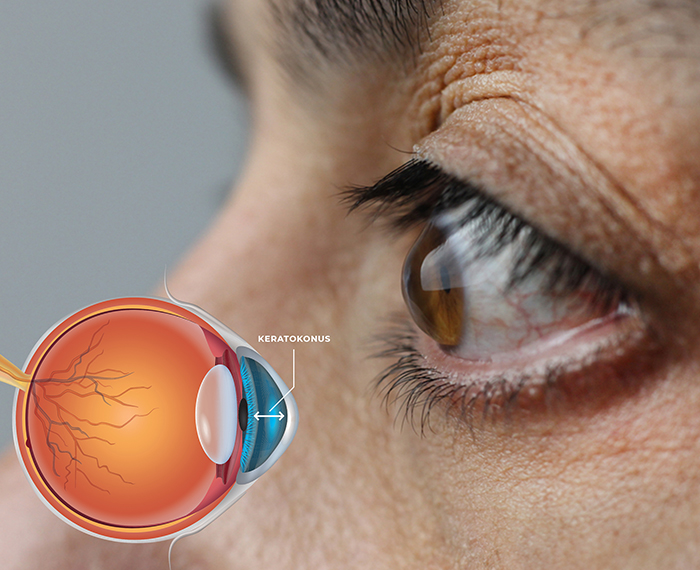
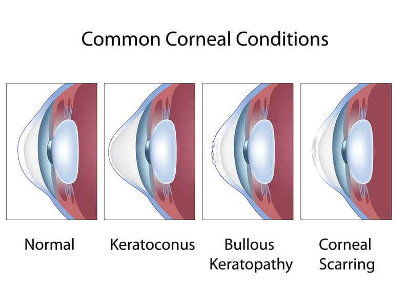
Keratoconus is a progressive eye disease with thinning and tapering of the anterior corneal layer of the eye, leading to irregular astigmatism and myopia leading to decreased visual function.
What are the symptoms?
The most common symptoms are increased myopia and astigmatism for a short period of time and consequently frequent eyeglasses change, poor vision despite eyeglasses, decreased night vision, increased light sensitivity and difficulty in reading.
Who is seen and why?
It is seen in both women and men without showing gender differences. In the etiology, recurrent trauma due to scratching and genetic factors were responsible. It is often seen in people with allergic eye disease such as atopic conjunctivitis and vernal conjunctivitis, which cause excessive itching in the eyes. It has also been reported to be associated with genetically inherited conditions such as retinis pigmentosa (chicken land disease), Down syndrome, and some connective tissue diseases. Today, the most accepted view is that keratoconus is a multifactorial disease and it is caused by environmental factors (such as eye scratching) in people with genetic predisposition.
What is the course of the disease?
Keratoconus usually begins in adolescence or in the twenties and progresses at a rate varying from person to person. This progress stops around 30-40 years and the disease becomes stable. Although the disease affects both eyes, it is quite asymmetric and often more prominent in one eye.
How is keratoconus diagnosed?
An experienced ophthalmologist can recognize advanced keratoconus patients with a standard eye examination. However, in suspicious cases, corneal topography and thickness examinations are made by special examination devices such as Orbscan and Pentacam. These special examination methods are also used to determine whether keratoconus is progressing and to evaluate the outcome of the treatments.
What are the treatment methods?
The first step in the treatment of keratoconus is to try to stop the progression if the disease is in progress. The rate of disease progression varies from person to person and progression is usually seen in young people. The examinations are repeated every 3 to 4 months to see if there is any progress. Other methods used to improve visual acuity in patients with progressive stopped keratoconus are intracorneal ring application, intraocular lens application and corneal transplantation.
Collagen crosslinking treatment
The only proven method to stop progress today is “crosslinking CC (CCL). However, for the CCL method to be applied safely, the diagnosis should not be late, that is, the cornea should not be too thin. In CCL method, ultraviolet light is applied to the eye after the eye is dripped with riboflavin drops. In this way, the hardness of the cornea is increased and strengthened by the photochemical reaction.
Contact Lenses
Patients with keratoconus who have poor vision with glasses may benefit from hard contact lenses or hybrid contact lenses (soft and hard combination) specially designed for keratoconus. Contact lenses are not suitable for advanced patients with very thin corneas.
Intra-Corneal Ring Method
In the ring method, semicircular prostheses made of polymethylmethacrylate or acrylic polymers are inserted into the tunnels prepared by femtosecond laser in the cornea. In order to apply the ring method, the progression of keratoconus must be stopped; therefore, pre-ring CCL treatment should be applied. In addition, in order to apply ring treatment, the cornea should not be too thin and not too steep. Corneal rings placed in the cornea in the appropriate eyes provide the best corrected visual acuity by flattening the cornea.
Intraocular lens application
In patients with progressive keratoconus who cannot benefit from contact lens and ring therapies, another treatment option to relieve myopia and astigmatism is to apply intraocular lens. These lenses are placed on the person’s own transparent lens or on the colored iris layer. In order to perform such an application, the patient must have sufficient corneal endothelial cell count.
Corneal transplantation
Corneal transplantation is usually used as a last resort in patients with advanced keratoconus who have been neglected and have very thin corneas. Approximately 20% of keratoconus patients go to corneal transplantation. Transplantations for the treatment of keratoconus are promising, with over 90% success in the first 10 years.